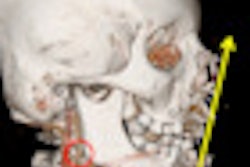
For the analysis of gunshot injuries using CT, 2D multiplanar reformatting (MPR) and 3D volume-rendering (VR) postprocessing software can provide valuable additional information to axial imaging, particularly in the examination of vessels and bone lesions, Italian researchers have reported.
"Ballistics analysis can be improved through 2D-MPR and 3D-VR reconstructions on the Cartesian axes, and nonconventional software such as radiotherapy planning software," noted Dr. Marco Matteoli, from the department of radiology at S. Andrea Hospital, La Sapienza University in Rome, and colleagues in a poster presentation at last month's RSNA 2013 meeting in Chicago.
Multidetector CT allows accurate determination of fireflight injuries, mainly in emergency and war settings, and permits the surgeons and anesthetists to plan their interventions more thoroughly and become aware of clinically occult injuries. The technique facilitates visualization in 2D or 3D by providing an isotropic volumetric dataset and creating reformatted images of excellent quality and significance, they explained.
Firearm use is a common method of suicide and homicide, particularly in the U.S. For instance, there are more than 1,000 firearm-associated fatalities a year in Los Angeles County alone, according to the authors. In penetrating injuries, the bullet enters into the body and does not exit, while in perforating injuries, the bullet enters and exits from the body.
Of great importance is the permanent wound cavity, which is the path taken by the bullet while crushing and effectively destroying the tissue. The cross-sectional area of this cavity is also equal to the bullet's cross-section at that same point modified by a shape factor derived from the bullet's configuration and the amount of resistance offered to it by the tissue, they stated.
Radial stretching of tissue occurs around the bullet's wound track, which momentarily leaves an empty space called the temporary wound cavity. The size of the temporary cavity is approximately proportional to the kinetic energy of the striking bullet and also the amount of resistance the tissue has to stress. Bullets, associated fragment, and blast debris may enter a vessel and embolize to the lungs or brain.
In their RSNA exhibit, the group provided detailed information for each region of the body about the reconstruction algorithms and tissues to observe, as shown in the table.
| Region to examine | Reconstruction algorithm | Tissues to observe |
| Head | Axial view | Brain parenchyma, ventricular lesion, middle line shift, bleeding |
| 2D MPR/MIP sagittal | Venous vessels, position of bullet, bone lesions | |
| 2D MPR/MIP coronal | Bone lesions, position of bullet | |
| 3D VR/3D pathway | Trajectory of bullet | |
| Face | Axial view | Orbit and eyes, facial nerves |
| 2D MPR/MIP sagittal | bone structures | |
| 2D MPR curved | Vessels | |
| 3D VR | Bone structures | |
| Neck | 2D MPR/MIP sagittal | Airways, spine |
| 2D MPR curved | Smooth tissues, vessels, airway | |
| 3D VR | Vessels, position of bullet | |
| Thorax | Axial view | Lung parenchyma, heart |
| 2D MPR/MIP sagittal | Airways, spine, thoracic aorta, diaphragm | |
| 2D MPR/MIP coronal | Airways, pulmonary vessels, heart | |
| 3D VR/3D pathway | Trajectory of bullet | |
| Abdomen | Axial view | Solid and hollow organs |
| 2D MPR/MIP sagittal | Abdominal aorta, spine | |
| 2D MPR/MIP coronal | Iliac and femorals, peripheral vessels | |
| 3D VR/3D pathway | Vessels, position of bullet, bone structures, trajectory of bullet | |
| Limbs | ||
| 2D MPR/MIP curved | Vessels, smooth tissues | |
| 3D VR | Bone structures, position of bullet |
"CT interpretation in standard assessment of axial sections can be improved in real time at the workstation through postprocessing software using reconstruction algorithms such as 2D-MPR imaging in sagittal, axial, coronal, or curved view, 2D multiplanar maximum intensity projection (MIP), and 3D-VR imaging," they wrote.
Bullet placement is one of the most important factors in achieving incapacitation. Tissue trauma consists of both the permanent wound cavity and also radial stretching of surrounding tissue, resulting in temporary cavitation. The extent of trauma depends on the type of bullet involved, the kinetic energy and mass of the bullet, the size of the organ or tissue, the presence or absence of anatomical constraints from adjacent structures, and the resistance of the tissue to strain, explained Matteoli et al.
Although surgical planning needs correct evaluation of solid or hollow organ lesions, vessel and nerve resections, diaphragm lesions and bone fractures, CT evaluation for solid organ injuries in abdominal or chest trauma does not usually require special reconstructions, and in most cases axial scanning is sufficient to define the extension of the splanchnic lesions to treat, they concluded.


