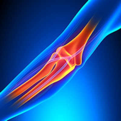
Elbow injuries are common in power and combat sports like boxing, judo, weightlifting and wrestling, but uncertainty surrounds the best imaging approach. Belgian researchers provided fresh analysis at the 2015 congress of the European Society of Musculoskeletal Radiology (ESSR) and collected a top award for their work.
For assessment of tendon pathology in the elbow, plain film and ultrasound are the first-choice modalities and are usually sufficient for the lateral, medial, and posterior compartments, but ultrasound evaluation of the anterior compartment is difficult, according to Dr. Milan Vansevenant, a radiologist at Ghent University Hospital and AZ Sint-Maarten in Mechelen, Belgium, and colleagues.
"Clinical examination is a prerequisite in the assessment of tendon pathology at the elbow," they noted. "MRI is useful in the case of refractory symptoms, confounding cases, for a global overview, and for differential diagnosis. The FABS (flexed elbow, abducted shoulder, forearm supinated) position is useful for the evaluation of the distal biceps tendon."
Elbow tendinosis is very frequent in daily practice and is often due to overuse or intensive sports activities, explained the researchers, whose e-poster was one of three top prize-winners selected by judges at the annual congress of the ESSR, held in York, U.K., in late June. Microtearing occurs during stress on a tendon, and if the stress is repetitive, there will be no full repair because of the hypovascular nature of a tendon and the repetitiveness of the stress with new microtearing, resulting in tendinosis and eventually macrotears.
Direct trauma is another cause of tendon injury and the mechanism of trauma differs along with the involved compartment. The therapeutic strategy depends on the involved compartment and the mechanism of injury, they added.
Imaging's precise role
In most cases, there is no need for imaging after a clinical examination and history, but in therapy-resistant or severe cases, or in the presence of confounding symptoms, imaging becomes mandatory and is particularly useful to aid preoperative planning, Vansevenant et al stated. Serious tendon pathology or associated abnormalities should not be overlooked.
Plain films are useful in initial workup to rule out occult fractures or stress fractures of the elbow (see table). Indirect signs of tendinosis are bone spur formation or soft-tissue calcifications at the insertion of the affected tendon. Plain films have a limited role in early disease, and although the same indirect signs can be seen on CT, CT is not preferred because of its greater radiation dose.
"Ultrasound is the first line examination in the evaluation of the different elbow compartments," they wrote. "The probe is placed in a longitudinal and axial plane on the axis of the tendons. Tendon thickening, hypoechogenicity, hyperemia, calcifications, and (partial) tears are seen. To evaluate hyperemia on echo-Doppler imaging, compression with the probe should be avoided so as not to compromise the blood flow. Hyperemia represents an angiofibroblastic reaction."
Elastography can be used to measure the stiffness of tissues, representing it on a color map. Healthy tendons are hard and have low compressibility, whereas in tendinopathy, tendons become softer and have high compressibility.
MRI is a second-line examination in the evaluation of elbow pain, and is excellent to show associated pathology and to rule out other causes of elbow pain than tendon pathologies, according to Vansevenant and colleagues. Normal tendons and ligaments are of uniform low-signal intensity on all imaging sequences, and tendon thickening, signal intensity changes of the tendon, or underlying bone marrow and tears of the tendon and/or adjacent ligaments can be seen in pathologic conditions.
They recommend a slice thickness of 3 mm and a high-resolution coil, and the preferred field of view is 10 cm to 14 cm. The patient should be put in the gantry in the so-called "superman" position, with the elbow in extension and supination. T2-weighted and proton density-weighted images with fat saturation are very useful to evaluate fluid, and T1-weighted images are used for structural evaluations.
In the lateral compartment, plain films can show bone spur formation or soft-tissue calcifications near the lateral epicondyle in advanced cases, while on MRI the tendons and ligaments of the lateral compartment are best evaluated on axial and coronal images. Also, thickening of the common extensor tendon, signal intensity changes, tears and associated tears can be efficiently evaluated on MRI.
In the anterior compartment, plain films usually have no added value caused by superposition. CT can be useful to reveal bone spur formation in the distal biceps tendon.
The authors use the FABS-position for assessment of the distal biceps tendon on MRI. In this position, the tendon loses its oblique course seen in the standard imaging position. The tendon is straight in a coronal and sagittal plane, resulting in an easier interpretation of the distal biceps tendon. Plain films of the elbow can show anchor placement after distal biceps tendon repair.
In the posterior compartment, plain films can show bone spur formation or soft-tissue calcifications near the olecranon in advanced cases. Ultrasound of the distal triceps tendon is performed with the elbow in flexion. In MRI, the triceps tendon is best evaluated in the sagittal and axial plane. After trauma or repetitive stress, avulsion fracture of the olecranon can occur. Olecranon bursitis is usually a straightforward diagnosis on ultrasound due to its location at the olecranon and in the subcutaneous soft tissue. It can show a fluid collection with usually hyperemia, and there may be some debris in the collection, Vansevenant et al concluded.


