Dear Advanced Visualization Insider,
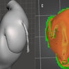
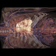
The integration of advanced visualization technologies into medicine has led to improvements in a wide range of clinical applications over the past several years. In particular, the tangible 3D view provided by patient-specific 3D-printed anatomical models has enhanced the visualization of complex anatomy and, in doing so, boosted the efficiency of challenging procedures.
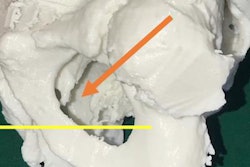
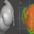
In a recent example published in the Turkish Journal of Urology, urologists from India demonstrated how 3D-printed pelvic models based on CT scans helped direct the course of treatment for patients with urethral injuries and also facilitated preoperative planning.
A separate study in Italy showed the benefits of using 3D printing to resolve a similarly difficult case involving complications in the upper gastrointestinal tract. The researchers concluded that a patient-specific 3D-printed model proved to be "fundamental" to safe surgical intervention.
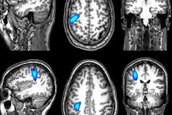
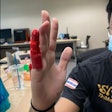
In other news, a group from the U.K. presented a prototype magnetoencephalography (MEG) scanner capable of capturing 3D brain images of adults and children while they perform simple tasks. The MEG helmet could pave the way for a better understanding of brain development and various neurological conditions, the group said.
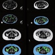
Finally, the recent European Society of Cardiovascular Radiology congress in Belgium had various presentations discussing the potential advantages of combining artificial intelligence (AI) and advanced visualization technologies to assist in clinical tasks. In one such presentation, researchers from the Utrecht University Medical Center in the Netherlands demonstrated the feasibility of quantifying body composition through automatic segmentation of 3D CT scans with AI.
For more recent news and research, head over to the Advanced Visualization Community at AuntMinnieEurope.com. As always, feel free to contact me with any topics you'd like to see covered in the coming days.
