When the doors of the ECR 2011 technical exhibition burst opened this morning, a host of surprises and innovations awaited congress attendees. Arguably the biggest highlight of all is the first PET/MR system ever displayed at the Austria Center, and it looks certain to attract massive attention and interest over the next four days.
A combination of MRI and PET has long been considered as the logical next step in the evolution of imaging modalities, but industry has struggled to deal with the formidable technical challenges of achieving a happy marriage between the two approaches, notably the difficulties of developing a PET detector capable of coping with the powerful static and dynamic magnetic fields generated by the MR coils. GE Healthcare made the first important move in developing the software needed to integrate data acquired through sequential scans made using the two modalities. Then Philips brought the two main items of hardware alongside each other, linked by a revolving table for easy patient transfer, and the company installed the first European system in a hospital in Geneva in April 2010. Now Siemens' engineers have successfully merged the two modalities in a single unit, dubbed the Biograph mMR, which the vendor plans to launch commercially later this year.
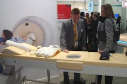
In the meantime, radiologists will be able to examine the prototype on display in the technical exhibition, and if they are lucky, they will get to talk to Dr. Markus Schwaiger and his colleagues from the Klinikum rechts der Isar at the Technical University in Munich. The group has been given the privilege of putting the new machine through its paces.
Dr. Alexander Drzezga, from the university's department of nuclear medicine, has been responsible for setting up the system, which was delivered in November 2010. It is currently being used to scan up to five patients a day, but that number is likely to grow as the Munich team explores the limits of the unit's clinical potential. "Many neurological conditions are suitable for evaluation with PET/MR, including neurodegenerative disorders, dementia, epilepsy, and brain tumors. With regard to evaluation of the cardiac system, combined imaging of PET and MR may also show diagnostic advantages, while inflammation and vascular conditions are also areas of interest," he explained.
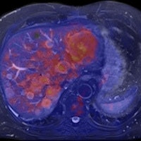
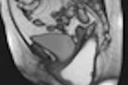
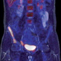 Left: PET tracer uptake in the liver can be combined with the time-varying enhancement of dynamic MR scans to visualize hepatic tumor characteristics. Right: MR and PET come together to support tumor staging. In this case, high-resolution MR provides a clear image of the pathology within the pelvic structure, while PET displays the hypermetabolism component of the lesion. Images courtesy of Siemens.
Drzezga believes that combining the two modalities offers a number of clinical advantages, not least in eliminating the need for separate diagnostic examinations. Furthermore, the exact anatomical registration of structural and functional/molecular information may improve allocation of suspect findings and improve image quality, for example by motion correction of regions of the body that do not remain rigid during examination. The Munich team will also be exploring how the performance of PET/MR compares with that of PET/CT. There is some evidence that the superior soft-tissue contrast achievable with the newer system will offer significant benefits, even before physicians consider the safety issues involved with any radiation-based imaging technology such as PET/CT, Drzezga suggested.
The Biograph mMR is based on the Verio 3-tesla MR system with a 70-cm bore that provides enough space to position the PET detector ring and its solid-state photodiodes. In getting the two modalities to work alongside each other, the hybrid molecular MR system can scan the whole body in as little as 30 minutes, compared with an hour or more needed for sequential examinations. The machine also incorporates Siemens' TIM (total imaging matrix) technology, which seamlessly integrates multiple coil elements and radiofrequency (RF) channels and can reduce examination times by up to 50%, according to the vendor.
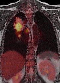
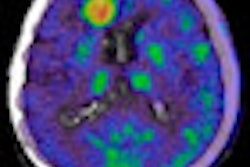
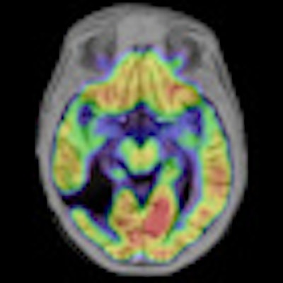
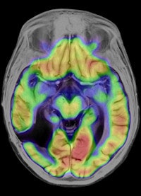 Left: To improve the precision of spatial registration, the Biograph mMR system collects MR and PET data simultaneously from a single frame of reference. The result is a combined MR and PET scan acquired at the same point in time and reflecting the same point in the physiologic processes such as respiration. Right: The benefits of MRI in the study of neurological diseases are well-known and established, and it can lead to a better understanding of neurological pathologies. Images courtesy of Siemens.
Philips has been working on its latest MR technology for almost as long as the search for a practical PET/MR hybrid. After an eight-year development project, the company is promoting its Ingenia 1.5- and 3-tesla systems, which it describes as the world's first digital broadband MR unit. This incorporates dStream architecture, which digitizes the signal directly in the coil. Vendors have been looking for ways to shorten the analogue part of the signal processing pathway because this offers the potential for reducing signal loss and noise. The Philips approach goes further by digitizing the signal within the coil itself and transporting it via a fiber-optic cable to the acquisition electronics contained in the scanner cabinet, explained Maurits Wolleswinkel, global lead for MR marketing.
 High-resolution MR image of the hip with spectral attenuated inversion-recovery (SPAIR) fat suppression. Image courtesy of Philips.
The company had to overcome huge challenges in miniaturizing the components to fit inside the coils and making them tough enough to cope with the hostile physical environment inside the machine, with its strong magnetic fields, eddy currents, and temperature changes. "The result offers a gain of up to 40% in the signal to noise ratio and a 30% improvement in throughput, and eliminates the need for coil/channel upgrades as the system is totally channel-independent," he stated. "So it is a significant step forward, especially in oncology-related applications. They become better, faster, and more robust. We can do a total liver examination, including contrast, in less than eight minutes, or a whole-body diffusion scan in 15 minutes."
 Two-station torso imaging with multitransmit. Image courtesy of Philips.
Improving the patient's experience when he or she is undergoing an MR examination has been the main focus of GE Healthcare's latest developments in this modality. This aspect can have a considerable impact on both the diagnostic accuracy of the procedure and on workflow within the department, noted Marie-Caroline du Réau, GE's European MR marketing manager. "If we can create a stress-free environment, the patient feels more comfortable and is less likely to fidget inside the machine and spoil the exam. So we can get a better quality image and make more efficient use of the technologist's time."
The main highlight of the company's offerings at ECR 2011 is a wide-bore 3-tesla MR system, the Discovery MR750w, which incorporates its proprietary ART (acoustic reduction technology) system for reducing scanner noise. GE is also showing its new Optima MR430s 1.5-tesla device, which is designed for examining musculoskeletal injuries of the extremities and can free up places on the departmental worklist for those patients needing more complex whole-body examinations.
Both these machines are commercially available and come equipped with the GEM suite, a set of receive-only RF surface coils. These can be used individually or combined to provide the desired anatomical coverage, including head to toe coverage. Overall, the range covers 98% of examination types. The GEM suite allows for independent selection of 160 coil-mode configurations, and the system will autoselect the configuration that best fits the selected region of interest, the vendor stated. Other key features include a total 205-cm scanning range, feet-first scanning in many cases, and design features to embrace patients of all shapes and sizes.
Originally published in ECR Today March 4, 2011.
Copyright © 2011 European Society of Radiology


